
.jpg)
Practice Innovations
Infrared thermography (IRT) applications in wound care continue to emerge1,2,3, and over more than traditional single-point temperature measurement (thermometry). IRT captures sounds of temperature readings within the image and can be used to create gradients and contrasts that reveal patterns that can be visually assessed to guide clinical decision-making. Compared to thermometry, this offers the greatest advantage to the method; however, training is required to interpret the patterns. This abstract reveals the critical steps to utilizing IRT within wound care practice and highlights key patterns to review in the IRT image beyond the absolute temperature or measured temperature difference between two points.
Methods:
Using a series of case studies, this presentation reviews key principles of thermographic imaging with a focus on visual inspection. A series of case images from various wound types (e.g., diabetic foot ulcer, pressure injury, venous leg ulcer, infected vs. Non-infected) to analyze different patterns that appear in thermographic images.
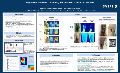
Results:
: Providing a structured process for collecting IRT images and a framework to review may aid wound care clinicians in recognizing patterns to inform clinical decision-making. Acquisition protocols are important for image standardization. Palette contrast selection is also important for highlighting focus areas. Typical patterns include cooler wound bases caused by evaporative heat loss from moisture and exudate. In contrast, asymmetrical thermal changes or high contrasts in temperature caused by large temperature differences can be critical indicators of underlying inflammation or infection that need to be recognized.
Discussion:
By understanding how to collect and interpret IRT images effectively, wound care clinicians can identify subtle changes in the wound status that might go unnoticed by only using single-point measurements or a temperature difference. Visual inspection of IRT images utilizes the thousands of temperature data points collected in the IRT image, providing a better picture of wound healing progress and risk.