Laboratory Research
(LR-014) Comparative Analysis of Amniotic Membrane Allografts Modulation of Keratinocyte Activity in Vitro
Friday, May 2, 2025
7:45 PM - 8:45 PM East Coast USA Time

Sarah Moreno, MS – MIMEDX Group, Inc.; Michelle Massee, BS; John Harper, PhD
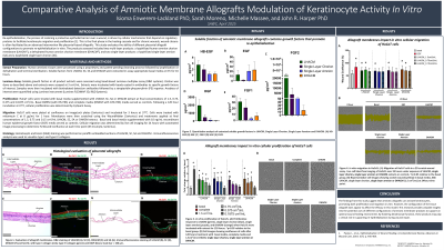
Introduction: Re-epithelialization, the process of restoring a protective epithelial barrier over a wound, is driven by cellular mechanisms that depend on regulatory proteins to facilitate keratinocyte migration and proliferation [1]. This is the final phase in the healing cascade and for chronic wounds, wound closure is often facilitated by an advanced intervention like placental-based allografts. This study evaluates the ability of different placental allograft configurations to promote re-epithelialization in vitro. The products assessed included two multi-layer products: a lyophilized human amnion chorion membrane (LHACM*), a dehydrated human amnion chorion membrane (DHACM#), and two single-layer products: a lyophilized single-layer amnion (SA) and a lyophilized single-layer chorion (SC).
Methods: Allografts were prepared using the PURION® process, which includes gentle cleansing, lyophilization or dehydration, and terminal sterilization. Histological analysis using hematoxylin and eosin (H&E) staining and immunofluorescence (IF) characterized structural and compositional differences. Multiplex Luminex assays quantified regulatory proteins related to epithelialization in allograft extracts. Subsequent analysis of the functional activity of these extracts was evaluated in migration and proliferation assays using HaCaT cells, an immortalized human keratinocyte cell line. All comparisons were performed using equivalent surface area to volume ratios to model clinical application.
Results: The structural composition of each allograft was assessed using H&E staining, allowing visualization of the physical differences of each product. Immunofluorescence staining identified collagen I and IV, providing additional information on allograft composition. Luminex assays showed allograft extracts contained factors associated with re-epithelialization. All products promoted cellular migration at the highest concentration tested. At the lowest concentration, only the multi-layered products (LHACM and DHACM) demonstrated accelerated scratch wound closure. Similarly, all products promoted keratinocyte proliferation at the highest concentration, however at the lowest concentration, only LHACM showed a significant effect.
Discussion: The findings from this study suggest that amniotic allografts can activate keratinocytes, promoting both proliferation and migration in vitro. However, the configuration of the tissue allograft does appear to affect the efficacy in this model. This research provides valuable insights into the potential uses of different configurations of amniotic membrane products to support an optimal wound healing environment. By fostering keratinocyte function, these products may play a critical role in supporting re-epithelialization during wound repair.

.jpg)