(CS-126) Martorell's Hypertensive Ischemic Leg Ulcer: A Case of Mistaken Identity
Friday, May 2, 2025
7:45 PM - 8:45 PM East Coast USA Time
Frank Aviles, PT; Anil Matta, MD, MPH
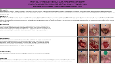
Introduction: 45 year old female presented to the wound care clinic with an atypical wound to the left medial lower extremity. The patient reported trauma at the etiology resulting in a small black bump that gradually worsened. Past medical history is positive for morbid obesity, severe OSA, and Htn. Social history is positive for smoking 1 ppd x 10 years. The wound presented with eschar and extremely painful. The rest of the history was benign.
Methods: Laboratory testing showed elevated inflammatory markers. Imaging was negative. Sharp debridement was performed with a punch biopsy to the margin. Pathology was nonspecific- concern for stasis dermatitis vs PG. The pt was started on high dose steroids. There was worsening pain, size and overall appearance. A wedge biopsy was performed and sent to dermatopathology. Results showed marked hyperplastic media of midsized arterioles with complete occlusion of lumen and calcium deposition, suggestive for Martorell's Hypertensive Ischemic Leg Ulcer.
Results: Steroids were rapidly tapered. The patient was started on calcium channel blockers. Extensive debridement was performed and NPWT and 4 layer compression was utilized until the wound was appropriate for a split thickness skin graft. The wound showed dramatic improvement with the above interventions with eventual healing.
Discussion: This case represents the importance of identifying Martorell's Hypertensive Ischemic Leg Ulcer and differentiating between Pyoderma Gangrenosum and Vasculitis as the treatments are completely divergent.

.jpg)