
.jpg)
Laboratory Research
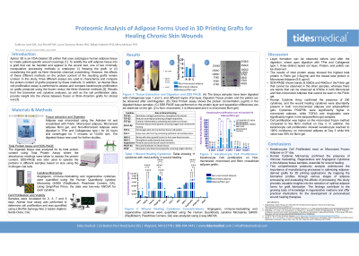
The application of 3D-printed adipose-derived grafts holds promise for the treatment of chronic skin wounds by promoting enhanced tissue regeneration. This study aims to map the proteomic profile of human adipose tissue at various stages of processing to identify key biomarkers that contribute to wound healing outcomes.
Methods: Using the microarray Q4000 assay, the proteomic content was analyzed across four adipose forms: raw, micronized, gelled, and frozen. The influence of different sterilization methods, including supercritical carbon dioxide (SrCO2) and electron beam (e-beam), on protein integrity was also evaluated. This proteomic mapping provides insight into how processing and sterilization impact the regenerative potential of adipose-derived grafts.
Results: Preliminary findings indicate significant differences in the total protein content and distribution among the different adipose forms, as visualized through colorimetric dye assays and SDS gel electrophoresis. The comparison between SrCO2 and e-beam sterilization demonstrated distinct proteomic patterns, suggesting that sterilization methods may play a critical role in preserving bioactive proteins. Heatmap data analysis revealed unique proteomic signatures at each stage of processing, highlighting variations in biomarker abundance that could impact the graft’s effectiveness in chronic wound healing applications.
Discussion: This comprehensive proteomic analysis underscores the importance of manufacturing processes in optimizing adipose-derived grafts for 3D printing applications. By mapping the biomarker profiles through various stages of adipose processing and evaluating the effects of sterilization methods, this study provides valuable insights into the selection of optimal adipose forms for graft fabrication. The findings contribute to the growing body of knowledge in regenerative medicine and offer practical implications for the development of personalized wound healing therapies.