
.jpg)
Clinical Research
Wound images can have errors in color, which may impede diagnosis and distort measurements.1,2,3 Color errors are caused by technical variability in smartphone camera algorithms and settings, as well as inconsistent photographer technique during image acquisition.2,3 Color calibration improves technical variability and reduces color errors by more than half,3 however it does not correct for errors related to photographer technique. Source capture optimization and alignment guides (SCOAG), as used for remote banking deposits, help reduce photographer technique inconsistency and standardize the image acquisition process.4 To assess the effectiveness of SCOAG, Delta E values of uncalibrated photos taken with SCOAG and then compared to the National Institute of Standards and Technology (NIST) values for smartphone photographs.
Methods:
A database of 9,070 clinical photos taken between May 12, 2023 – October 13, 2023, by over 100 photographers from 12 Wound Care facilities using a variety of (Apple) smartphones were compared. A color chart (TRUE-See Systems) with target colors and QR code activated SCOAG was physically placed next to the wound during each image acquisition. The photos were assessed using The International Commission on Illumination standard Delta E 2000 measurement of color difference and compared to the National Institute of Standards and Technology (NIST) reported values for smartphone.
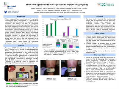
Results: The use of SCOAG improved image color 58.8% (12.7) over the NIST Baseline. Post calibration the SCOAG image color improved 73.1% (15.8) over the NIST baseline and 43.1% (4.4) over NIST calibration baseline
Discussion: This data clearly illustrates that implementing SCOAG can effectively standardize wound image acquisition and significantly enhance color quality. By operationalizing and harmonizing photo acquisition alongside color calibration, we can greatly boost the clinical relevance and reliability of images, streamline regulatory review processes, and drive innovation forward