Laboratory Research
(LR-040) Understanding Kinetics of Rapidly Vascularizing Composite Collagen Dermal Templates (CCDT)

Introduction: Reconstitution of skin anatomy following full-thickness skin loss can be achieved using dermal regeneration templates (DRTs) in combination with split-thickness skin grafts (STSGs). However, due to the limited rate of cellular infiltration and vascularization of currently available DRTs, reconstruction typically involves a two-step procedure spaced over several weeks. Our group previously demonstrated that a CCDT consisting of a collagen-based 3D structure with a microarchitecture maximizing differential density promotes enhanced cell and vascular invasion, and that a CCDT of 1.5 mm thickness could be placed with a STSG, resulting in a healed wound with reconstituted neodermal layer in a single-step procedure. Here, a follow-up study explores the underlying mechanisms behind these observations.
Methods:
Methods: CCDTs were prepared using established protocols. Female Yucatan pigs received 3x3 cm full-thickness skin wounds on their dorsum. Wounds were treated with either CCDT or a market-leading DRT (L-DRT), followed by application of standard dressings. Healing was assessed at three time points (3, 7, and 10 days post-wound creation). Wounds (N=5/group/timepoint) were harvested and analyzed.
Results:
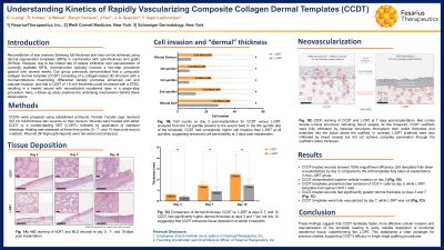
Results: By day 3 of the study, CCDT-treated wounds demonstrated 100% engraftment efficiency (5/5 templates) versus 0% (0/5 templates) in the L-DRT group. Additionally, the CCDT group presented superior cellular invasion on day 3, relative to the L-DRT (Fig. 1B). Moreover, by day 3, CCDT templates presented clear evidence of CD31+ cells within the templates, whereas the L-DRT demonstrated sparse individual CD31+ cells on the template/wound interface. This resulted in fully vascularized CCDT templates by day 7, while the L-DRT template achieved only 80% vascularization (Fig. 1A). Assessment of the newly formed dermal tissue revealed significantly greater dermal thickness in CCDT-treated wounds on days 3 and 7 as well as day 10 (Fig. 1C).
Discussion:
Conclusions: These findings suggest that CCDT facilitates faster, more effective cellular invasion and vascularization of the template, leading to early, reliable deposition of functional neodermal tissue, outperforming the L-DRT. This establishes a clear paradigm for previous studies, supporting CCDT’s efficacy in single stage grafting procedures.

.jpg)