(CS-103) Thermal Imaging Analysis of Diseased Skin in Hidradenitis Suppurativa Patients
using Forward-looking Infrared (FLIR) Technology: A Pilot Study
Friday, May 2, 2025
7:45 PM - 8:45 PM East Coast USA Time
Seanna Yang, BS – Medical Student, Department of Dermatology, Tulane University School of Medicine; Samantha Morin, BA,BS – Medical Student, Department of Surgery, Division of Plastic and Reconstructive Surgery, Tulane University School of Medicine; Laurel Adams, BS – Medical Assistant, Department of Surgery, Division of Plastic and Reconstructive Surgery, Tulane University School of Medicine; Claire Hines, BA – Medical Student, Tulane University School of Medicine; Andrea Murina, MD – Professor, Department of Dermatology, Tulane University School of Medicine; Harry Dao, MD – Associate Professor and Chair of Dermatology Department, Department of Dermatology, Loma Linda University; Abigail Chaffin, MD, FACS, CWSP, MAPWCA – Professor and Division Chief, Department of Surgery, Division of Plastic and Reconstructive Surgery, Tulane University School of Medicine
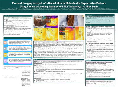
Introduction: Hidradenitis suppurativa (HS) is a chronic inflammatory skin disorder which affects the folliculopilosebacous unit. The exact pathophysiology of HS is not yet fully understood; however, it has been linked to upregulation of inflammatory cytokines, leading researchers to agree that the HS cycle of inflammation begins before visible manifestations. Given the known presence of subclinical HS disease, we aimed to investigate forward-looking infrared (FLIR) thermal imaging as a non-invasive tool to identify and map active inflammation through temperature analysis. FLIR technology has the potential to help refine surgical resection boundaries not visible to the naked eye. This pilot study evaluates the utility of FLIR imaging in mapping diseased skin in patients with HS to optimize surgical planning and improve outcomes.
Methods: Patients with HS undergoing surgical excision were imaged pre- and post-operatively using standard photographs and FLIR thermal imaging. To preserve consistency, patients were acclimated to operating room temperature before imaging. FLIR data were analyzed to denote diseased tissue from healthy tissue. Diseased tissue was considered areas with elevated temperatures indicating greater inflammation. Postoperatively, patients were monitored for persistent disease and complications.
Results: This case series included two patients who underwent surgical excision of HS. FLIR imaging identified high-temperature areas the extended beyond the surgical borders drawn around disease and inflammation visible to the naked eye, involving multiple anatomical sites (inguinal region, medial thigh, anterior vulva, and posterior labia). In Patient 1 (inguinal and left thigh resection), HS disease was successfully excised. Post-operative healing was complicated by mild cellulitis and wound dehiscence which did not require operative intervention. Patient 2 (vulvar and labial resection) also had complete disease excision and healed without complications.
Discussion: This pilot study establishes a foundation for larger-scale investigations and highlights the potential role of FLIR thermal imaging in enhancing surgical planning for patients with HS. The methods employed in this study for analyzing infrared images provide a framework for future research into the application of thermal mapping to optimize surgical interventions in HS.

.jpg)