(LR-015) Tri-layer Amniotic Membrane Allografts Promotes Re-epithelialization in Vitro and Ex Vivo
Friday, May 2, 2025
7:45 PM - 8:45 PM East Coast USA Time
Sarah Moreno, MS – MIMEDX Group, Inc.; Michelle Massee, BS; John Harper, PhD
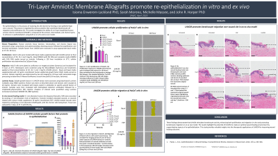
Introduction: Re-epithelialization is the process of restoring the skin barrier by forming a new epithelial layer over a wound. This process is regulated by various proteins that coordinate keratinocyte cell migration and proliferation [1]. This study investigates the ability of a tri-layer lyophilized human amnion chorion membrane (LHACM*), composed of the amnion, intermediate, and chorion layers, to enhance re-epithelialization using both an in vitro and ex vivo model.
Methods: LHACM was prepared using the PURION® process, which involves gentle cleansing, followed by lyophilization and terminal sterilization. Multiplex Luminex assays were employed to detect and quantify factors responsible for promoting re-epithelialization in LHACM extract. Effects on cellular proliferation and migration were evaluated in HaCaT cells, an immortalized human keratinocyte cell line. Furthermore, the effects of LHACM on re-epithelialization were evaluated using a human ex vivo skin model. In this model, 11 mm-diameter human skin biopsies were wounded by excising a 2 mm-diameter circle of epidermis. The wounds were treated by overlaying a 4 mm-diameter LHACM graft, with the chorion side facing down.
Results: The LHACM extract contains soluble factors that promote keratinocyte migration and proliferation. Treatment with LHACM extract significantly enhanced HaCaT cell proliferation in vitro and accelerated wound closure, indicating increased cellular migration. In the ex vivo wound model, LHACM grafts facilitated the migration of activated keratinocytes into the wound bed, further supporting its role in re-epithelialization.
Discussion: These findings demonstrate that LHACM stimulates keratinocyte activity, enhancing both proliferation and migration in vitro and promoting keratinocyte migration in an ex vivo model. The results highlight the potential of LHACM to create an optimal wound healing environment by supporting key aspects of re-epithelialization. This study provides valuable insights into the therapeutic applications of LHACM for improving wound healing outcomes.

.jpg)