(LR-026) In Situ Colorimetric Nanofiber Membrane for Bacterial Detection at Wound Sites: Validation of Accuracy and Visual Feasibility
Friday, May 2, 2025
7:45 PM - 8:45 PM East Coast USA Time
Farinaz Jonidi Shariatzadeh, Ph.D. Candidate – Research Assistant, Biomaterials Synthesis and Surface Engineering Lab, Department of Biosystems Engineering, Faculty of Agriculture and Food Science, University of Manitoba; Sarvesh Logsetty, MD, FRCSC FACS – Director Manitoba Firefighters Burn Unit, Professor Departments of Surgery, Psychiatry, Children’s Health, Rady Faculty of Health Sciences, President Canadian Burn Association, University of Manitoba; Maurice Haff, ME, MBA – CTO & Adjunct Professor, College of Business, ParaNano Wound Care, LLC & University of Central Oklahoma; Song Liu, Ph.D., P.Eng – Professor, Biomaterials Synthesis and Surface Engineering Lab, Department of Biosystems Engineering, Faculty of Agricultural and Food Sciences, University of Manitoba
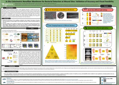
Introduction: Detecting infections in wound care remains a significant challenge, particularly in hospitals and home care settings. Current clinical methods, such as swabbing and culturing, are time-intensive, require specialized training, and delay critical intervention. By the time physical symptoms appear, bacterial loads may exceed critical thresholds, increasing the risk of systemic infections. To address this, we developed an in situ colorimetric nanofiber membrane capable of real-time detection of bacteria at concentrations below infection levels. Unlike existing technologies requiring expensive equipment, our membrane offers a cost-effective, user-friendly solution that changes color from yellow to green in response to bacterial lipase, allowing detection with the naked eye.
Methods: The membrane was fabricated using electrospinning, combining a core solution of polyurethane with a shell solution containing polyurethane, polyvinylpyrrolidone, hemicyanine dye, citric acid, and Tween 80. We evaluated its response to bacterial lipase and its specificity by exposing it to solutions of salts (containing Na, Mg, K, Fe) and proteins (bovine serum albumin and fetal bovine serum rich in different enzymes and proteins). To assess usability across various settings, we tested the membrane under different lighting conditions (daylight, cool light, and biosafety cabinet light) and angles (45º and straight). Color changes were quantitatively analyzed using spectrophotometry, recording CIELAB and HEX# values as reference controls.
Results: The biosensor demonstrated high specificity to bacterial lipase, with no false positives observed in the presence of salts or proteins. Bacterial tests confirmed that the color change was easily distinguishable under diverse lighting conditions, with only minor variations between the colors under different light but significant difference to control without bacteria. These findings validate the biosensor’s reliability and practicality for real-time bacterial detection.
Discussion: This in situ nanofibrous colorimetric biosensor offers a promising solution for early bacterial detection in wounds, reducing risks and financial burdens on patients and healthcare systems. Its simplicity, accuracy, and adaptability make it suitable for various applications, including hospitals, home care, and battlefield settings, empowering even untrained users to monitor wound infections effectively before its progress to systemic infection or damaging adjunct tissues.

.jpg)