(CS-133) Effects of Human Cryopreserved Adipose Tissue Allograft Implantation on Tissue Oxygenation in Diabetic Neuropathic Patents with Plantar Ulcers: Insights from Multispectral Near-infrared Spectroscopy Imaging
Friday, May 2, 2025
7:45 PM - 8:45 PM East Coast USA Time
Introduction: Plantar ulcers, common in diabetic neuropathic individuals with fat pad atrophy, account for nearly half of all foot ulcers and are a leading cause of limb amputations, with rates exceeding 80%. High plantar pressure due to fat pad loss plays a key role in ulcer formation, poor healing, and recurrence. Human cryopreserved adipose tissue allograft (hCAT) has emerged as a promising approach for managing fat pad atrophy.1 This study evaluates the impact of hCAT implantation on tissue oxygenation in diabetic neuropathic patients with plantar ulcers.
Methods: This retrospective case series included 3 patients (2 males, 1 female) with multiple comorbidities, including diabetes, and plantar ulcers that failed to respond to previous treatments. The ulcers varied in size (0.6–3.4 cm²). A comprehensive wound management approach included the application of 3.0 mL of hCAT to address fat pad defects. Tissue oxygenation and visual light images were captured using a mobile near-infrared spectroscopy imaging device**.2 Imaging was performed at various time points, from 7 to 108 days prior to hCAT implantation, continuing up to 181 days post-implantation. Follow-up intervals were tailored to individual cases. The primary outcomes included the rate of wound closure and kinetics of tissue oxygenation after hCAT implantation.
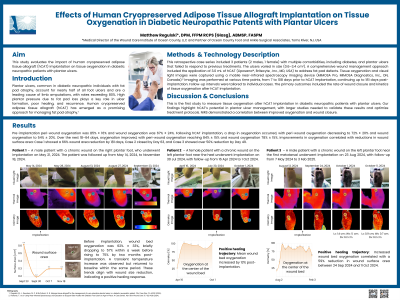
Results: Pre-implantation peri-wound oxygenation was 85% ± 16% and wound oxygenation was 67% ± 24%. Following hCAT implantation, a drop in oxygenation occurred, with peri-wound oxygenation decreasing to 72% ± 20% and wound oxygenation to 54% ± 20%. Over the next 18–64 days, oxygenation improved, with peri-wound oxygenation reaching 84% ± 15% and wound oxygenation 78% ± 15%. Improvements in oxygenation correlated with reductions in wound surface area: Case 1 showed a 66% wound area reduction by 181 days, Case 2 closed by Day 63, and Case 3 showed over 50% reduction by Day 49. Longer-term follow-up is ongoing.
Discussion: This is the first study to measure tissue oxygenation after hCAT implantation in diabetic neuropathic patients with plantar ulcers. Our findings highlight hCAT’s potential in plantar ulcer management, with larger studies needed to validate these results and optimize treatment protocols.

.jpg)