
.jpg)
Case Series/Study
Peripheral vascular disease (PVD) is a global issue affecting roughly 200 million people. This disease comes secondary to atherosclerosis of vessels and can lead to complications such as lower extremity ulceration and amputation [1]. Following a lower extremity amputation, mortality rates increase from 39% to 80% in five years [2]. A multidisciplinary approach in conjunction with dermal substitutes have shown success in wound healing and limb salvage [3]. This case study demonstrates the efficacy of a bilayer biodegradable synthetic matrix (BBSM) and monolayer biodegradable synthetic matrix (MBSM) to heal a complex PVD ulcer secondarily.
Methods:
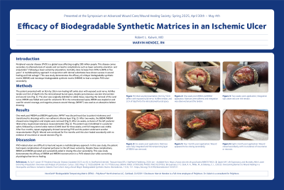
The patient presented with an 8 cm by 10 cm non-healing left ankle ulcer with exposed sural nerve, Achilles tendon and 2 cm of depth into the retrocalcaneal bursal space, despite percutaneous vascular intervention and wound care. The ulcer was surgically debrided to viable tissue, requiring the removal of the sural nerve. MBSM was folded and used for volumetric fill in the retrocalcaneal space, BBSM was stapled on and used for wound coverage, and negative pressure wound therapy (NPWT) was used as an absorptive bolster dressing.
Results:
NPWT was discontinued due to patient intolerance and transitioned to dressings with a non-adherent silicone layer. After two weeks, the BBSM/MBSM showed some integration and staples were removed. After six weeks, occlusion of the left posterior tibial artery required percutaneous revascularization. The patient was immobilized in a posterior splint, followed by a control ankle motion (CAM) boot for three weeks, until full integration was visible. After four months, repeat angiography showed worsening PVD and the patient underwent another revascularization. Wound care continued for five months until the ulcer healed secondarily with no evidence of recurrence or wound necrosis.
Discussion:
PVD related ulcers are difficult to heal and require a multidisciplinary approach. In this case study, the patient had repeat complications of impaired perfusion to the left lower extremity. Despite these complications, BBSM and MBSM persisted and successfully aided in closing this ulcer secondarily. This case study demonstrates the efficacy of BBSM and MBSM reconstruction in a PVD related ulcer while overcoming physiological barriers to healing.