
.jpg)
Clinical Research
The adoption of minimally invasive techniques, such as foam sclerotherapy and radiofrequency ablation (RFA), for treating chronic venous insufficiency has increased significantly over the past decade.¹ While duplex ultrasonography (DUS) remains the gold standard for diagnosing venous insufficiency and evaluating treatment efficacy,² it is time-consuming and resource-intensive. Near-infrared spectroscopy (NIRS) imaging, a non-invasive modality, offers spatial information on tissue oxygenation (StO₂) and has the potential to streamline evaluations. This study aims to explore the utility of NIRS imaging in assessing the efficacy of minimally invasive treatments.
Methods:
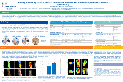
A quasi-experimental pre-posttest study was conducted on 14 patients treated for chronic venous insufficiency and venous leg ulcers with either foam sclerotherapy or RFA between November 2022 and February 2024. Tissue oxygenation and skin surface temperature were measured using an FDA-cleared handheld multispectral NIRS and infrared (IR) temperature imaging device (MIMOSA Pro, MIMOSA Diagnostics Inc., Toronto, Canada).³,⁴ NIRS images were taken pre- and post-treatment from the treatment side and the contralateral (control) side, including various anatomical locations: the dorsum and plantar aspects of the foot, medial and lateral leg, and wound area (if present). Statistical analyses, including paired t-tests, were used to evaluate pre- and post-treatment differences.
Results:
Successful vein closure after RFA and sclerotherapy was confirmed in all 14 cases (100%) using postoperative DUS, with no reopening observed during the follow-up period. Post-treatment NIRS data showed an average 20% increase in mean StO₂ on the plantar surface for 13 cases (93%), while one case (7%) showed no clinically significant change. Before treatment, the mean StO₂ on the treatment side was 53%, compared to 60% on the control side (Figure 1). After treatment, the mean StO₂ on the treatment side increased to 73%, representing a statistically significant improvement (p < 0.05).
Discussion:
Near-infrared spectroscopy imaging provides a reliable, non-invasive method for real-time monitoring of tissue oxygenation. By visualizing microcirculation changes, NIRS enables clinicians to detect treatment success or failure earlier, facilitating timely interventions. While the small sample size limits generalizability, these findings highlight the potential of NIRS imaging as a valuable clinical tool for optimizing vascular treatment outcomes. Future research with larger cohorts is recommended to validate these results.